Monday, February 10, 2020

Sciatica/Lumbar Radiculitis Care At Ocala Family Medical Center


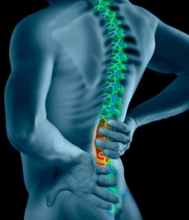
A pinched nerve in the spine, also known as radiculopathy, can lead to a variety of uncomfortable symptoms, including numbness, pain, and weakness. Compression and irritation of the sciatic nerve, or sciatica refers to pain that may affect any of the five spinal nerve roots that give rise to each sciatic nerve. Both sciatic nerves travel from the low back through the buttock and down the back of the leg and foot.


 Along with pain down the length of the sciatic nerve there are other symptoms of sciatica may include difficulty with movement, numbness, muscle weakness, and tingling. Symptoms are usually unilateral, meaning they occur in only one sciatic nerve at a time, although sometimes symptoms are perceived on both sides but symptoms are usually unilateral, meaning they occur in only one sciatic nerve at a time.

Radiculopathy may occur for no apparent reason or as the result of an injury. Most likely to experience radiculopathy are those individuals aged 30 to 50 years old, and the effects will be felt in the cervical and lumbar spine areas. Directly along the course of a specific spinal nerve root, radicular pain radiates into the lower extremity which includes the thigh, calf, and occasionally the foot. The most common symptom of radicular pain is sciatica, which is pain that radiates along the sciatic nerve. This discomfort is felt down the back of the thigh and calf into the foot. One of the most common forms of pain caused by compression of a spinal nerve in the low back, sciatica is often caused by compression of the lower spinal nerve roots.


 Leg pain is typically much worse than the low back pain with this condition while the specific areas of the leg and/or foot that are affected depends on which nerve in the low back is affected. Radicular pain into the front of the thigh and the shin is caused by compression of higher lumbar nerve roots.

Compression of lumbar or sacral nerves, or by compression of the sciatic nerve itself is the general cause of sciatica. A bulging spinal disk, deterioration and misalignment of the vertebrae, and a herniated intervertebral disc in the spine can be the cause of this nerve compression. Degenerative disc disease is a common cause of these changes. Tears in the annulus fibrosis, the tough outer skin of the intervertebral discs,involves breakdown of the vertebrae and may allow the soft inner nucleus pulposus (pulp) to protrude and push against spinal nerves. This can cause severe pain and numbness and this nerve compression is a most common cause of sciatica.


Spinal stenosis, or progressive narrowing of the spinal canal causing spinal nerve or spinal cord compression, is another cause of sciatica. Bone spurs, herniated discs, which decrease available space for the spinal cord, inflammation and spondylolisthesis can all be the cause of this narrowing. This condition also pinches and irritates lumbar and sacral spinal nerves, and this in turn affects sciatic nerves. Persistent pain in the low back and lower extremities, which may include sciatica, often results from spinal stenosis. Other symptoms of the disorder are decreased sensation in the lower extremities, decreased physical activity and difficulty walking. Spinal stenosis patients often present with bilateral sciatica, which means that both sciatic nerves are affected.

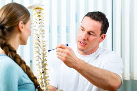
Doctors may diagnose radiculopathy using MRI and CT scans as well as radiologic imaging with X-ray in addition to a physical exam and a review of symptoms. Some physicians may employ electrical impulse testing called electromyography or EMG which can test nerve function.


In the beginning the treatment of sciatica focuses on lowering the inflammation that is causing the patient's symptoms. Initial treatments might include taking muscle relaxers and non-steroidal anti-inflammatory drugs (NSAIDs) as well as physical resting. To treat more stubborn cases of inflammation your doctor may prescribe oral steroids, which have more severe side effects but are proven effective. Radiculopathy usually is treatable without surgery. Doctors may recommend certain medications, including oral corticosteroids or injectable steroids depending on the severity of the radiculopathy. These may include narcotic pain medications.


A course of nonsurgical treatment such as selective spinal injections, physical therapy, medications, a soft cervical collar and ice and heat applications should be conducted for six to eight weeks. Decompressive surgery, such as discectomy/microdiscectomy, and/or laminectomy , may be recommended if nonsurgical treatment does not alleviate the pain. For 85% to 90% of patients this type of surgery typically provides relief of radicular pain/leg pain.


Physical Medicine & Rehabilitation


team! Dr. Cruz is board certified in Physical Medicine &Rehabilitation
(PM&R), with fellowship training in Interventional Spine and Sports
Medicine. Matt Ota is a Board Certified Physician Assistant with
extensive knowledge of musculoskeletal disorders and injuries.


(located at OFMC Plaza)
2135 SW 19th Avenue Road

Wellness Direct: 352-368-1340
OFMC Main: 352-237-4133
Fax:      352-873-4581

Tuesday, December 31, 2019

Peripheral Neuropathy Treatment At Ocala Family Medical Center


The affliction known as peripheral neuropathy is not a single disease. The name is a general term for a series of disorders that result from damage to the body’s peripheral nervous system. Two parts comprise the body’s nervous system - our central nervous system includes the spinal cord and the brain, while the peripheral nervous system connects the nerves running from the brain and spinal cord to the rest of the body.

When nerves are damaged or destroyed and can not send messages from the brain and spinal cord to the muscles, skin and other parts of the body then peripheral neuropathy occurs. When this damage happens numbness and pain in the arms, hands, legs, and feet may occur. Multiple nerves (polyneuropathy) can be affected or peripheral neuropathy can attack only one nerve or nerve group (mononeuropathy) at a time. Usually mononeuropathy is the result of damage to a single nerve or nerve group by inflammation, trauma, injury, local compression, or prolonged pressure.


 Being an intricate network, our peripheral nerves connect the brain and spinal cord to the muscles, skin, and internal organs. Peripheral nerves coming out of the spinal cord, peripheral nerves are arranged along lines in the body called dermatomes. Damage to a nerve will typically affect one or more dermatomes, which can be tracked to specific areas of the body. The resulting injury to these nerves interrupts communication between the brain and other parts of the body. This damage can impair muscle movement, prevent normal sensation in the arms and legs, and cause pain. As well it can also affect other areas of your body.

Traumatic injuries, infections, metabolic problems, inherited causes and exposure to toxins can all be causes of peripheral neuropathy. Diabetes is one of the most common causes. Be aware that peripheral neuropathy can affect one nerve (mononeuropathy), two or more nerves in different areas (multiple mononeuropathy) or many nerves (polyneuropathy). An example of mononeuropathy is Carpal tunnel syndrome, and most people with peripheral neuropathy have polyneuropathy.


 Generally speaking the signs and symptoms of peripheral neuropathy might include localized numbing, a gradual onset of numbness, prickling or tingling in your feet or hands, which can spread upward into your legs and arms. You might also experience a sharp, jabbing, throbbing or burning pain and an extreme sensitivity to touch. Some patients report pain during activities that should not cause pain, such as pain in your feet when putting weight on them or when they are under a blanket. Sometimes there is lack of coordination and falling and muscle weakness. Signs and symptoms if autonomic nerves are affected might include changes in blood pressure, causing dizziness or lightheadedness, bowel, bladder or digestive problems, excessive sweating or not being able to sweat and heat intolerance.

Your physician will review your medical history and conduct a physical exam and neurological evaluation before diagnosing peripheral neuropathy. This neurological evaluation, which consists of a number of simple and painless tests is usually performed to diagnose peripheral neuropathy. You may encounter other tests that can determine what type of peripheral neuropathy you have depending on your symptoms and outcome of the neurological evaluation.


 It will be easier for your doctor to understand your condition if you are prepared to discuss your symptoms in detail with them. Most likely your doctor will ask you to describe your symptoms, when you experience them, how long the episodes last and the amount of discomfort or pain you experience. Make your doctor very aware of your condition by being specific about the tingling, numbness, weakness or other symptoms you are experiencing.

Keep in mind that your doctor may also ask you general health questions that may seem unrelated to your symptoms, but are very important. Such questions could be about whether or not you feel faint, nauseated or tired, and your doctor may also ask if your bladder control and sexual function are normal. Usually you will also be asked if you are taking medications or suffering from any other illnesses.

In every case your treatment is based on treating the underlying disorder, for example If diabetes is the cause, making certain that the blood glucose is controlled is important. Correcting the deficiency is the treatment if a vitamin deficiency is causing the problem. It has been discovered that many treatments can bring relief and help you return to your regular activities, and sometimes a combination of treatments works best.


 Sometimes medications are the temporary solution as over-the-counter pain medications like acetaminophen (Tylenol) and nonsteroidal anti-inflammatory drugs, such as aspirin and ibuprofen, can be very helpful in controlling moderate pain. These drugs can affect your liver or stomach function if you take them in excess so it is most important to avoid using them for an extended period, especially if you drink alcohol regularly.

To control the pain of this condition many prescription pain medications can also help. In this group are narcotics, antidepressants and some antiepileptic medicines. Your doctor may prescribe other helpful prescription medicines including seizure medications, such as gabapentin or pregabalin, tramadol or corticosteroid injections.

Another treatment that may be right for you can include surgery. You might need surgery to reduce the pressure if you have neuropathies caused by pressure on nerves, such as pressure from tumors. And finally the key for you might include physical therapy. This can help improve your movements if you have muscle weakness. Medical equipment might also be required, aids such as a wheelchair, hand or foot braces, a cane, a walker, or a rollator.


Physical Medicine & Rehabilitation


team! Dr. Cruz is board certified in Physical Medicine &Rehabilitation
(PM&R), with fellowship training in Interventional Spine and Sports
Medicine. Matt Ota is a Board Certified Physician Assistant with
extensive knowledge of musculoskeletal disorders and injuries. 


(located at OFMC Plaza)
2135 SW 19the Avenue Road

Wellness Direct: 352-368-1340
OFMC Main: 352-237-4133
Fax:      352-873-4581

Friday, November 29, 2019

Photaging Dermatology Care At OcalaFamily Medical Center


We all need our time in the sun in a poetic, philosophical manner. But we have to be very careful when we encounter the brilliant light of our closest star. Ultraviolet (UV) light damages your skin every time you go outdoors without sun protection or use a tanning bed. This damage builds up with time and you see changes to your skin, which can make you look years older than you naturally would. Sunlight damage changes to skin can include wrinkles,age spots, loose skin, spider veins and a blotchy or ruddy complexion.

For these changes “Photoaging” is the medical term although you may also hear the terms “premature aging” or “sun-damaged skin.” As the damage builds, You may see deep lines or dry, scaly patches called actinic keratoses (AKs). Be aware that an AK is a pre-cancerous skin growth, as sun damage can also lead to skin cancer. In some cases an AK turns into a type of skin cancer called squamous cell carcinoma. With the effects of excessive sunlight, while much of the damage is permanent treatment can reduce some signs of sun damage that are making you look older.


A great barrier against the sun’s ultraviolet (UV) rays can be provided by clothing. Protective clothing's protection is consistent over time and does not wear off like sunscreen does. High-tech protection and breathability are offered by many new fabrics. High neck, long sleeves, and pants work best as the more skin you cover the better, Also keep in mind that a hat with a wide brim all the way around at least three inches or more is best because it helps shade your neck, eyes, ears, and face. You should as well always wear UV-blocking sunglasses to protect your eyes and the skin around them.


During the hours of 10 am to 4 pm the sun is strongest and during this time it is best to minimize sun exposure. It would be best to plan your outdoor activities for the early morning or late afternoon. While it can certainly feel good to spend time in the sunshine it can take a toll on your skin because the sun gives off ultraviolet (UV) light that damages your skin and causes sunburn. You can add years to your looks over time as these rays can lead to wrinkles, dark spots, and other problem areas. UV exposure is the reason behind 80% of your skin’s aging, research shows.


Invisible rays of ultraviolet light are given off by the sun. Ultraviolet B (UVB) rays are short, high-energy wavelengths that are absorbed by the epidermis, the outermost layer of the skin. The skin responds to UVB rays by producing chemicals called inflammatory mediators when you burn and these seep down into the dermis, the skin's middle layer. The dermis is irritated as these chemicals distress the tiny blood vessels in the dermis. These small vessels then swell and create the surface redness we see in a sunburn.


 Be aware that the UVB rays affect the genetic material of the epidermis and this causes the damage that may lead to skin cancer. The immune system can be affected and other UVB rays can interfere with the skin's ability to repair itself. Furthermore UVB radiation attacks the skin's melanocytes, which are the pigment cells. The reaction of the melanocytes is to step up production of melanin and send melanasomes to the skin's surface to act as a filter against the sun's rays and this can actually damage the DNA of the pigment cells. Genetic damage of this sort can cause both freckling and the mottled brown of age spots. Unfortunately it also contributes to the development of melanoma and other skin cancers.


 Sun damaged skin in some cases can be repaired with laser treatments, including Q-switched, Photorejuvenation, and Fraxel. Fraxel tackles both the brown spots and fine wrinkles associated with excessive sun exposure and is an excellent treatment for sun damaged skin. Redness and brown spots on the face caused by skin sun damage can be tackled with little or no downtime by Photorejuvenation treatments. This non-invasive procedure that makes your face look more youthful by removing and reversing signs of rosacea, sun damage, and reducing pores or acne spots is also called Photofacial, Fotofacial or IPL. It remains one of the most popular treatments due to its low downtime and effective results.


Ben Treen, M.D. and Amber Starling APRN-C make up OFMC’s Dermatology team! They specialize in the diagnosis, treatment and prevention of skin diseases and cancers. Our in-house pathology lab is also located at the Dermatology, Aesthetics & Women's Health Center.


Dermatology Direct: 352-368-1350
OFMC Main: 352-237-4133
Fax:      352-873-4581


Tuesday, October 29, 2019

Ulnar Neuropathy Treatment At Ocala Family Medical Center


Running all the way from your shoulder to your little finger your ulnar nerve manages the muscles that allow you to make fine movements with your fingers. The ulnar nerve also controls some of the muscles of your forearm that allow you to grip things tightly. The ulnar nerve is not protected by muscle or bone throughout its course unlike most of your other nerves. Being near the surface of your skin means that ulnar nerve injuries aren’t uncommon and the ulnar nerve is what creates the shock-like sensation when you hit the funny bone in your elbow.

If you damage your ulnar nerve you may lose sensation and have muscle weakness in your hand. This condition is known as ulnar nerve palsy or ulnar neuropathy and it can affect your ability to make fine movements and perform many routine tasks. Ulnar nerve palsy can cause muscle wasting, or atrophy, that makes the hand look like a claw in severe cases. When this happens surgery is sometimes necessary to correct the condition.


 Probably the most common site of pain in an ulnar neuropathy is the elbow area, while many people complain of sensory changes in the fourth and fifth digits. Less commonly, an individual actually notices that the unusual sensations are mainly in the medial side of the ring finger (fourth digit). In other cases the third digit is also involved, especially on the ulnar (medial) side. The resulting sensations can be a feeling of numbness or a tingling, and pain rarely occurs in the hand. Pain complaints tend to be more common in the arm, up to and including the elbow area.

Ulnar nerve compression at the elbow is called "cubital tunnel syndrome" as the most common place for compression of the nerve is behind the inside part of the elbow. Numbness and tingling in the hand and fingers are common symptoms of cubital tunnel syndrome. Symptoms can be managed with conservative treatments like changes in activities and bracing in most cases. Your doctor may recommend surgery if conservative methods do not improve your symptoms, or if the nerve compression is causing muscle weakness or damage in your hand. It may be harder to complete tasks that were once easy since ulnar nerve palsy can make it difficult to work with your hands.


 To confirm a diagnosis of ulnar neuropathy a physical examination is usually all that is needed. Your doctor may order an electromyogram or nerve conduction studies if the symptoms are severe and require more aggressive treatment First your doctor will ask you about your symptoms and examine you. If your symptoms began after an injury to your hand make sure to tell your doctor as this can help him determine potential causes of your condition more easily. Your health care practitioner will want to know what makes your symptoms worse or better, how the injury occurred, and how long symptoms have been present.

Your doctor will assess the condition of your hand and evaluate how well you can move your fingers during the exam and in addition to a physical examination, testing can include X-rays, nerve conduction tests, imaging tests, such as a CT scan or MRI and blood tests. These examinations and diagnostic tests will measure nerve function in the ulnar nerve and help detect swelling. This will help your doctor to localize the area of the nerve that is not functioning properly, while a nerve conduction study can help determine the severity of the dysfunction.


 Similar treatments exist for cubital tunnel syndrome and ulnar tunnel syndrome. To relieve any inflammation that might be compressing the nerve is the first aim and this involves a combination of supportive splinting, rest, and proper mechanics. Ibuprofen or another pain reliever may also be prescribed. Steroid injections are generally not used because there is a risk of damage to the nerve even though steroids such as cortisone are very effective anti-inflammatory medicines. Doctors sometimes prescribe bracing or splinting, a padded brace or split to wear at night to keep your elbow in a straight position. Performing nerve gliding exercises may help some patients, many doctors think. These are exercises to help the ulnar nerve slide through the cubital tunnel at the elbow and the Guyon's canal at the wrist. In some cases these workouts can improve symptoms as they may also help prevent stiffness in the arm and wrist.


 Your doctor may recommend a surgical procedure that involves tendon transfer if he determines that the nerve is not going to restore its normal function. During a tendon transfer surgery, A functioning tendon is moved from its original bone attachment to a new one during a tendon transfer surgery and this can help to allow you to perform routine activities once again. Nerves heal slowly but the results of surgery are generally good, though full restoration of wrist and hand function can take months. You may still have a loss of sensation and movement in your hands even after surgery.


Physical Medicine & Rehabilitation


team! Dr. Cruz is board certified in Physical Medicine &Rehabilitation
(PM&R), with fellowship training in Interventional Spine and Sports
Medicine. Matt Ota is a Board Certified Physician Assistant with
extensive knowledge of musculoskeletal disorders and injuries.


(located at OFMC Plaza)
2135 SW 19the Avenue Road

Wellness Direct: 352-368-1340
OFMC Main: 352-237-4133
Fax:      352-873-4581

Wednesday, September 25, 2019

Diabetes Treatments At Ocala Family Medical Center


Glucose (blood sugar) is our brain's main source of fuel and is also an important source of energy for the cells that make up our muscles and tissues. The disease diabetes mellitus refers to a group of disorders that affect how our body uses glucose. An excess of sugar in our blood can lead to serious health problems no matter what type of diabetes we have.


Depending on the underlying cause, diabetes varies by type. Life-altering chronic diabetes conditions include type 1 diabetes and type 2 diabetes. There are possibly reversible diabetes conditions which include prediabetes, which can happen when your blood sugar levels are higher than normal, but not high enough to be classified as diabetes. There is also gestational diabetes which can happen during pregnancy but may resolve after the birth of the child.

Patients with type 2 diabetes are said to have insulin resistance and type 2 diabetes is a lifelong disease that prevents your body from using insulin the right way. Type 2 is the most common type of diabetes and there are about 27 million people in the U.S. with type 2. It is estimated that about 8 million people who have type 2 diabetes do not know it as the symptoms of it can be so mild you do not notice them.


 Some symptoms of type 2 diabetes include yeast infections that keep coming back, wounds that do not heal, feeling worn out, tingling or numbness in your hands or feet, being cranky a lot, being very thirsty, peeing a lot and blurry vision. Men with diabetes may have a decreased sex drive, erectile dysfunction (ED), and poor muscle strength in addition to the general symptoms of diabetes. Some women with diabetes can also have symptoms such as urinary tract infections, yeast infections, and dry, itchy skin.

By way of comparison the exact cause of type 1 diabetes is not known, though we do know that your immune system — which normally fights harmful bacteria or viruses — attacks and destroys your insulin-producing cells in the pancreas. When that happens it leaves you with little or no insulin and instead of being transported into your cells, sugar builds up in your bloodstream. While weight is not believed to be a factor in type 1 diabetes,type 1 is thought to be caused by a combination of genetic susceptibility and environmental factors, though exactly what those factors are is still unclear.


 It is of the utmost importance to get medical treatment for these conditions as untreated high blood sugar from diabetes can damage your nerves, eyes, kidneys, and other organs. Diabetes may also cause recurring infections because elevated glucose levels make it harder for the body to heal. Time is a real factor as the higher your blood sugar is and the longer you live with it the greater your risk for complications.

The treatment for diabetes depends on many factors and diet and exercise can help some people manage type 2 diabetes, but If lifestyle changes are not enough to lower your blood sugar, you will need to take medication. Different medications are used by doctors and some of these drugs are taken by mouth while others are available as injections. For type 1 diabetes the main treatment is insulin as insulin replaces the hormone your body is not able to produce. Thiazolidinediones help insulin work better while Alpha-glucosidase inhibitors slow your body’s breakdown of sugars and starchy foods.


You cannot treat diabetes, a serious disease, on your own. A diabetes treatment plan that is right for you and that you can understand can be provided by your doctor. It is probable that you will also need other health care professionals on your diabetes treatment team, including a nutritionist,foot doctor, eye doctor, and a diabetes specialist (called an endocrinologist). A combination of medications, exercise, and diet will help your treatment for diabetes as well as keeping close watch over your blood sugar levels and keeping them at a goal set by your doctor.


Family and Internal Medicine


Board Certified Primary Care providers are trained to care for you through all stages of life. Focused on building an on-going relationship, they help you stay well, treat you when you are sick and educate you on prevention and wellness to keep you and your family healthy.


Friday, August 30, 2019

Spinal Stenosis Remedies At Ocala Family Medical Center


The narrowing of the spaces within your spine which can put pressure on the nerves that travel through there is known as spinal stenosis. This malady occurs most often in the lower back and the neck region. Usually spinal stenosis is caused by wear-and-tear changes in the spine related to osteoarthritis. Doctors may recommend surgery to create additional space for the spinal cord or nerves in severe cases of spinal stenosis.


 While some sufferers may experience pain, tingling, numbness and muscle weakness that can worsen over time, some people with spinal stenosis may not have symptoms. A number of people are born with a small spinal canal and this is called “congenital stenosis”. In most cases,though, spinal narrowing is due to age-related changes that take place over time, and this is called “acquired spinal stenosis.”


 Your risk of developing spinal stenosis increases if you are female, were born with a narrow spinal canal, are 50 years old or older or if you have had a previous injury or surgery of the spine. There are certain medical conditions which can cause spinal stenosis including Paget’s Disease, spinal tumors, inflammatory spondyloarthritis and osteoarthritis, as well as bony spurs that form as we age.


 A rheumatologist will ask about your symptoms and medical history. If he suspects spinal stenosis he will do a physical exam and some symptoms he will look for include numbness, weakness, cramping or pain in the legs, thighs or feet that makes it hard to walk, as well as pain that goes down the leg. He may also want to know about any abnormal bowel and/or bladder function and loss of sexual function. There may even be, in severe cases, partial or complete leg paralysis. This is considered a medical emergency and he would likely instruct you to get to an emergency room as quickly as possible.


The location of the stenosis and the severity of your signs and symptoms will determine your treatment for spinal stenosis. Consult with your doctor about the treatment that is best for your situation. In the event that your symptoms are mild or you are not experiencing any your doctor may monitor your condition with regular follow-up appointments. Your health care provider may offer some self-care tips that you can do at home. Further treatments may be required if these do not help, in such a case your doctor may recommend medications or physical therapy.

If other treatments have not helped surgery may be an option. A number of recent studies have found that surgery produces better results than non-surgical treatment in the short term. Although surgery may bring some relief, it will not cure spinal stenosis or osteoarthritis and it is possible that your symptoms may recur.


 Medications may be needed to treat your condition, and your doctor may prescribe pain relievers. Ibuprofen (Advil, Motrin IB, others) and pain medications such as naproxen (Aleve, others) and acetaminophen (Tylenol, others) may be used temporarily to ease the discomfort of spinal stenosis. There is little evidence of benefit from long-term use of these medicines, so they are typically recommended for a short time only. Daily doses of tricyclic antidepressants, such as amitriptyline, can help ease chronic pain. Habit-forming medications that contain codeine-related drugs such as oxycodone (Oxycontin, Roxicodone) and hydrocodone (Norco, Vicodin) may be useful for short-term pain relief, but these opioids may also be considered cautiously for long-term treatment. Or your doctor may prescribe anti-seizure drugs, such as gabapentin (Neurontin) and pregabalin (Lyrica), which are used to reduce pain caused by damaged nerves.


 In an effort to reduce pain it is common for people who have spinal stenosis to become less active. However, this can lead to muscle weakness, which can result in more pain. The services of a physical therapist may be called for. Your physical therapist can teach you exercises that may help you improve your balance, maintain the flexibility and stability of your spine and build up your strength and endurance. A physical therapist can help you build and maintain strength in the muscles of your arms and upper legs, which will improve your balance and ability to walk. This could also help you bend and move about, as well as control pain.


Location


Wellness Direct: 352-368-1340
OFMC Main: 352-237-4133
Fax:      352-873-4581

Sunday, July 28, 2019

Treatment For Facet Joint Syndrome At Ocala Family Medical Center


Facet joint syndrome is the pain at the joint between two vertebrae in your spine, another term for facet joint syndrome is osteoarthritis. The joints in your spine that make your back flexible and enable you to bend and twist are the facet joints.The nerves in your back exit your spinal cord through these joints on their way to other parts of your body. Normal healthy facet joints have cartilage, which allows your vertebrae to move smoothly against each other with no grinding. All your joints are lubricated with synovial fluid for additional protection against wear and tear.


 Another term for facet joint syndrome is facet arthropathy. Progressive and degenerative changes in the spine may cause body weight to shift unevenly to the facet joints. The resulting extra stress causes wear and tear on the joint and changes it over time. Through this process the joint capsule thins and smooth cartilage breaks down and becomes irregular. Bone spurs may also form. These changes make it difficult for the joint to move fluidly and it reacts by becoming inflamed and irritated, similar to arthritis of the knee joint. As a result this irritated joint sends pain signals to the brain via small nerves in the capsule called medial branch sensory nerves. When this happens the muscles in the area can stiffen and spasm.


 Until an event triggers symptoms facet joint degeneration can be painless. Indicators that a person’s pain is coming from the facet joints may include a diffuse, dull ache in the low back directly over the spine that can spread to the buttocks. It can be felt in the shoulders and back of the skull, as well as in the neck. Twisting backwards or bending sideways towards the affected joint will cause pain. Periods of inactivity or standing may worsen the pain. Sitting, leaning forward, or changing positions, activities that take the weight off the joint, may ease the pain. A patient's facet joint symptoms can also mimic the pain of a disc herniation. In the event that bone spurs form and press on the spinal nerves, pain may be felt down the arms or legs. The pain may come in periodic flare-ups, or it may be chronic.


Cartilage in our joints wears down as we get older. Spine conditions that change the way the facet joints align and move can cause pain, events such as an injury, repetitive movements, obesity, and poor posture. The deterioration of a vertebral disc can begin changes in the facet joints. Over time when the load of the body weight shifts to the facet joint, the cartilage breaks down, the joint space narrows, and the bones rub together, leading to complications. Those prone to arthritis, both men and women, may be affected by facet joint syndrome. Usually it targets those between the ages of 40 and 70 and can also develop in people who have had a spine injury.

A complete history and physical exam usually begins the diagnosis of facet joint syndrome. Other diagnostic tests may be ordered by your doctor as well. To determine whether there are abnormalities in your spine, X-rays may be recommended. Sometimes a CT scan can show more detail about your facet joint surfaces. Your doctor may recommend a CT scan to get a better look if the X-rays suggest something may be affecting your facet joints. Your doctor may order a bone scan which can be useful in determining whether your facet joints are inflamed. In most cases an inflamed facet joint will show up as a hot spot on a bone scan.


 There is evidence that exercise, lifestyle changes and careful management of your back pain can contribute to better quality of life even though facet joint arthritis cannot be reversed. Your doctor may recommend injections, medications or surgery if conservative therapies fail to help you manage and control the pain. To prevent painful episodes using correct posture and keeping your spine in alignment are important things you can do. You can reduce the load on the facet joints and alleviate pain by losing weight and you might benefit further by making adjustments to your daily standing, sitting, and sleeping habits.


 Your doctor will likely recommend physical therapy to treat your symptoms once a diagnosis of facet joint syndrome has been confirmed. Well-rounded rehabilitation programs assist in improving your mobility and strength, helping you do your daily activities with greater ease and ability, and calming pain and inflammation. To decrease blood flow to the affected area and reduce swelling, physical therapy may also include the use of ice. To treat muscle spasms ultrasound and electrostimulation may also be used. Exercises will help you regain joint mobility, flexibility, and strength When you're feeling better and muscle stretching and massage may also be helpful.

In most cases you can heal faster through physical therapy, and exercise is very helpful for a painful facet joint. To strengthen and stretch your lower back, leg, and stomach muscles physical therapists will work with you. These specialists can instruct you on proper lifting and walking techniques. You must follow through, though...a physical therapist may show you strengthening and stretching exercises, but it is your responsibility to follow them.


 In some cases patients could require topical patches, oral anti-inflammatory medications or creams, salves or mechanical bracing. For muscle spasms, sometimes muscle relaxers are prescribed. A minimally invasive procedure that involves an injection of a corticosteroid and an analgesic-numbing agent into the painful joint is a facet joint injection, and steroids can reduce the swelling and inflammation of the nerves.


team! Dr. Cruz is board certified in Physical Medicine &Rehabilitation
(PM&R), with fellowship training in Interventional Spine and Sports
Medicine. Matt Ota is a Board Certified Physician Assistant with
extensive knowledge of musculoskeletal disorders and injuries.


Wellness Direct: 352-368-1340
OFMC Main: 352-237-4133
Fax:      352-873-4581